WinnMed is home to one of the area’s most comprehensive and advanced diagnostic imaging departments. State-of-the-art diagnostic radiology services include:
At WinnMed, patients benefit from the expertise of a full-time, board-certified radiologist, as well as skilled and compassionate registered technologists.
If your doctor orders a radiology examination, request a referral to WinnMed. We gladly provide radiology services to patients of all area health care providers.
An X-ray is a quick, painless exam that produces images of the solid structures inside your body, including bones and internal organs such as your lungs, heart and abdomen. A radiologic technologist takes the X-rays. You may experience discomfort during the exam depending on the position the technologist asks you to maintain. They will do their best to make it as painless as possible, while also ensuring they obtain quality images. Please tell your technologist if you experiencing any pain. Please inform the technologist if you are pregnant or could be pregnant prior to the test.
The examination is usually completed within 15-20 minutes. The X-rays are read by a radiologist and your doctor will discuss the results of the X-ray with you.
Are X-rays dangerous?
How do I find out my results?
My pain is on the right side. Why are you taking an X-ray of my left side?
Will my cell phone be OK in the X-ray room?
Can I get an X-ray if I am pregnant?
Fluoroscopy is a method of testing that uses real-time X-rays in order to assist the radiologist in viewing the digestive tract or joints. A special X-ray machine, called a fluoroscopy machine, is used. A radiologist performs the test with the assistance of a radiologic technologist. WinnMed offers the following tests:
GI stands for gastrointestinal, more commonly known as the digestive system. An Upper GI Series is an exam of the upper portion of your digestive system, including the esophagus, stomach and small intestine. Please inform the technologist if you are pregnant or could be pregnant prior to the exam. Expectant mothers, or women who think they may be pregnant, should not have an Upper GI Series.
An Upper GI Series is performed as an outpatient procedure. A radiologic technologist will position you next to the fluoroscope. You will be asked to swallow a small cup of liquid barium or similar substance. The radiologist, using the fluoroscope, will capture images of the liquid as it flows into your digestive system. You may be asked to change positions throughout the exam.
The examination is usually completed within 20-30 minutes and you can resume normal activity and diet directly following the exam. Your doctor will discuss the results with you.
Will I be under a form of anesthesia for an Upper GI Series?
Do you put a tube down my throat to look at my esophagus?
How long will the test take?
A barium enema is a special X-ray of the lower digestive tract, colon or large intestine. Since standard X-ray does not highlight soft tissue, a liquid barium solution is given by enema to make the colon and rectum visible to X-ray. A radiologist performs the test with the assistance of a radiologic technologist. Your doctor will discuss the results with you.
Please inform the technologist if you are pregnant or could be pregnant prior to the exam.
Barium enema is performed as an outpatient procedure. You will be asked to lie down on a table and a radiologic technologist will position you next to the fluoroscope. The technologist will insert a tube into your rectum. A liquid barium solution will be gently given by enema and x-rays will be captured as the solution outlines your colon. While the liquid barium solution fills your colon, you may experience mild cramping and/or abdominal discomfort. You may be asked to change positions during the exam. Following the exam, you will be asked to use the restroom to empty the barium from your colon.
Will I be under a form of anesthesia for a barium enema?
How long will the test take?
Ultrasound is an examination technique used to make still and live video pictures of the soft tissue structures of the body, such as the heart, digestive, reproductive or urinary tracts. Ultrasound is used to detect changes in appearance of organs, tissues and vessels or detect abnormal masses, such as tumors. Ultrasound is also used to monitor fetal growth and determine the age, weight and position of the fetus in the uterus.
Ultrasound is performed as an outpatient procedure. An ultrasound technologist (called a sonographer) will apply a clear, water-based gel to the area of your body to be examined. The sonogapher will guide a hand-held instrument, called a transducer, across the area being examined. Throughout the examination, the sonographer will capture images of the area for a radiologist to interpret. Your doctor will discuss the results of the ultrasound with you.
In some ultrasound studies, the transducer is attached to a probe and inserted into a natural opening in the body. WinnMed offers the:
Depending on the area to be examined, your doctor may provide special instructions to prepare for the exam. The procedure is painless with no short- or long-term side effects. Depending on the exam, an ultrasound will typically last 30-60 minutes; some up to 120 minutes.
When will I receive my results?
Is radiation used during an ultrasound?
WinnMed Radiology offers advanced breast cancer screening and diagnostic services, including 3D mammography (including mammograms for women with implants), ultrasound, ultrasound-guided breast biopsies and surgical biopsies in a convenient, comforting environment.
The American Cancer Society suggests:
Bone Densitometry, also known as bone density or DEXA, is a non-invasive scan of your bone mineral density or bone mass. It is a simple, painless exam that delivers approximately one-quarter to one-tenth the radiation that occurs during an ordinary chest X-ray.
People with porous bones, or osteoporosis, have higher risk for bone fracture. Bone densitometry provides information about your bones that, when compared to people whose age, sex and ethic background are similar to yours, can predict your potential for bone fractures. The most common risk factors for osteoporosis are:
Before your exam, a radiologic technologist may ask you to change into a gown. You will be asked to lie on your back on the examining table. The technologist will guide an imaging camera above your body that will move and take pictures. It is very important that you lie still while images are being taken so that the images are clear.
Please inform the technologist if you are pregnant or could be pregnant prior to the exam.
The exam takes approximately 15 minutes to perform. A radiologist will interpret the bone densitometry images and your doctor will discuss the results of the scan with you.
How often do I need a bone density test?
Why do you only scan my back and hips?
Is this radiation?
How can technologists be in the room without protection?
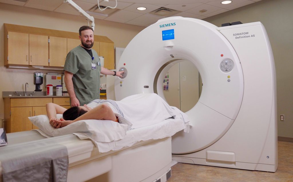
CT scan is short for Computed Tomography Scan, and is sometimes called CAT scanning. It is a noninvasive medical test that helps a radiologist study the inner workings of your body. CT scans of internal organs, bones, soft tissue and blood vessels reveal more details than regular X-ray exams. CT scans are particularly helpful in diagnosing and monitoring diseases of the central nervous system (brain and spine), as well as the chest and abdomen.
At WinnMed, we provide state-of-the-art technology with a 128-slice CT scanner. This means your doctor will have more images with more details to diagnose your illness or injury. The CT technology means our patients have shorter exam times, low dose exposure to radiation, and the larger machine helps to reduce anxiety for patients with claustrophobia and improves comfort for patients up to 676 pound.
Before your exam, a radiologic technologist will ask you to change into a gown and remove any jewelry. You will be asked to lie on the examining table, usually on your back. Depending on the area of the body to be examined, you may need a contrast agent to help produce clear images. Most contrast agents are injected into your body through a small IV needle. During the injection you may experience a metallic taste in your mouth and a hot, flushed feeling throughout your body. Please inform the technologist if you are allergic to iodine or have had a previous contrast reaction during a CT scan.
Once the exam begins, the table will move in and out of the CT machine opening, which is shaped like a donut. While the machine is running and images are being taken, you will hear a variety of whirring and clicking noises. Only the area of your body being examined will remain inside the scanner. It is very important that you lie still while images are being taken, so that images are clear. You may be asked to hold your breath during parts of the exam.
The technologist will monitor the machine from a computer in an adjacent room. He or she will be able to see, hear and communicate with you at all times. You will be able to talk with the technologist through an intercom system.
Please inform the technologist if you are pregnant or could be pregnant prior to the exam.
The exam is painless and can take from 10 minutes to one hour to perform. A radiologist will interpret the CT scan and your doctor will discuss the results of the scan with you.
When will I get my test results?
Does a CT scan hurt?
How long does a CT scan take?
If your doctor recommends an MRI, request a referral to WinnMed Radiology. We provide MRI exams daily using the latest in MRI technology. We serve everyone in our region, and will send the results to the doctor of your choosing.
Magnetic Resonance Imaging is more commonly referred to as MRI. MRI is a non-invasive procedure used to gather information about your body without involving radiation.
The MRI machine is a large magnet with a round opening. Using the magnet and radio waves, signals are sent to a computer that makes an image of the inside of the body appear on a screen.
MRI has been useful in diagnosing disorders of the central nervous system, joints, abdominal and pelvic organs, and circulatory system.
WinnMed has an in-house MRI machine. With patient convenience in mind, we provide scheduled MRI exams Monday – Friday.
Due to the magnetic properties of MRI, it is essential that we know about all metallic devices that may be present inside your body. They include pacemakers and/or pacewires, embedded shrapnel, aneurysm clips, cochlear stimulating devices, and artificial valves surgically affixed to your heart. You may be asked to have an X-ray of your eyes if you have a history of metal welding or grinding to ensure there is no metal present in your eyes. A radiologic technologist will review an MRI safety screening questionnaire with you prior to your scan to ensure you are sae to enter the MRI suite.
You will be asked to remove all loose or foreign metal objects such as jewelry, watches, dentures, credit cards and hairpins.
WinnMed’s MRI has a wide-bore magnet for added comfort during these studies. If you have concerns about lying in a small, enclosed space, mild sedation can help you comfortably complete the exam. Please discuss these concerns with your doctor prior to the exam to obtain the necessary medications.
Before your exam, a radiologic technologist will ask you to change into a gown and remove all loose or foreign metal objects such as jewelry, watches, dentures, credit cards and hairpins. Prior to entering the exam room, the technologist will screen your body with a metal detector to ensure your safety. You will be asked to lie on the examining table, usually on your back. You will be required to lie still during the MRI scan, and the technologist will do their best to get you as comfortable as possible. Depending on the area of the body to be examined, you may need a contrast agent to help enhance the images. Most contrast agents are injected into your body through a small IV needle.
Once the exam begins, the table will move inside a large circular opening that houses the magnet. The magnet is permanently open on both ends for patient comfort. While the machine is running and images are being taken, you will hear a variety of knocking and buzzing noises, which may become quite loud. To help reduce the sounds you will be provided ear plugs or you can select the type of music you would like to listen to throughout the exam.
The technologist will monitor you and your images through a large window the entire time. He or she will be able to see, hear and communicate with you at all times through a two-way intercom system. The technologist will also provide you with an alarm button to alert the technologist of any discomfort or concern you may experience at any point during the MRI exam.
If you are pregnant, MRI is not recommended. Please inform the radiologic technologist if you are pregnant or suspect you may be pregnant.
In general, most exams take between 45 minutes and 1 hour, but can take up to 2 hours to complete. A radiologist will interpret the MRI and your doctor will discuss the results of the scan with you.
Can I see my images?
When will I get my results?
How long does the test take?
A nuclear medicine scan is a safe, painless exam to help diagnosis abnormalities of internal organs and systems, especially abnormalities of the bone, gallbladder, heart, liver or thyroid.
The scan requires the use of a small amount of radioactive material, which is introduced into your system orally or with an IV injection. As the radioactive material travels to the area to be examined, a machine called a gamma camera makes detailed images of the area.
WinnMed offers nuclear medicine scans through a mobile service provided by Mayo Clinic.
A specialized technologist will provide an oral or IV injected dose of radioactive material. After the injection, you may be asked to leave and come back for the imaging in approximately 2.5–3.5 hours.
In most cases, you will not have to change into a gown for a nuclear medicine scan. You will be asked to lie on the examining table, usually on your back. You may be asked to change position as the technologist rotates the camera. It is very important that you lie still when asked by the technologist – sometimes up to 15 minutes at a time – so that images are clear.
Please inform the technologist if you are pregnant or could be pregnant prior to the exam.
The exam may take several hours to perform. A nuclear medicine physician will interpret the images and your doctor will discuss the results of the scan with you.
Is the radioactive material safe?
How long will the radioactive material stay in my body?